


Magnetic resonance imaging
MAGNETOM Aera 1.5T is a high-field magnetic resonance tomograph equipped with tunnel aperture of 70 cm which is comfortable for a patient during the procedure. MAGNETOM Aera 1.5T utilizes Tim + Dot + Trueform technologies. Tim technology allows for a combination of more than 204 elements of various rolls and selectable scanning area without repositioning of the patient, while Dot technology automatically selects the optimal scanning protocol for each subject. TrueForm forms cylindrical field of magnet uniformity to increase diagnostic value of images. The field is even throughout the scanning area, which allows for the increased investigational region and it helps to obtain high-quality images. Altogether, these methods help to the accuracy of diagnosis and improve comfort for patients under investigation.
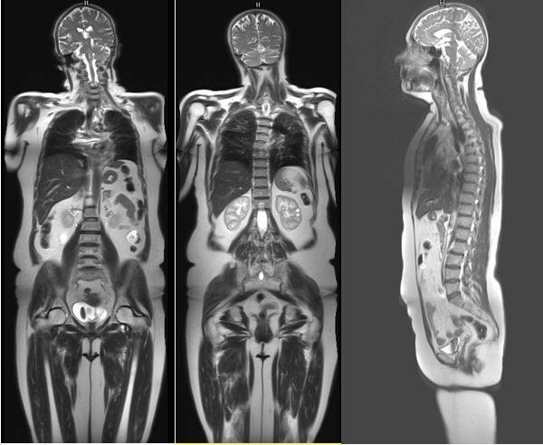
Figure 1 MRI of the whole body: scannning using Tim + Dot + Trueform technology (the investigation was performed in RRPC “Cardiology”)
Capacities of tomograph are enhanced due to the use of novel scanning sequencing and visualization analysis including heart function programme analysis; flow volume/velocity programme analysis, highly-specific myocardial tissue innovation analysis (Т1- Т2- Т2*- mapping); myocardial viability analysis adapted to use quantitative and volumetric assessment of fibrosis areas; myocardial perfusion analysis adapted to use visual and semi quantitative assessment;
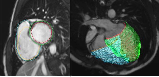
Figure 2 Heart MRI: funtional analysis (the investigation was performed in RRPC “Cardiology”)
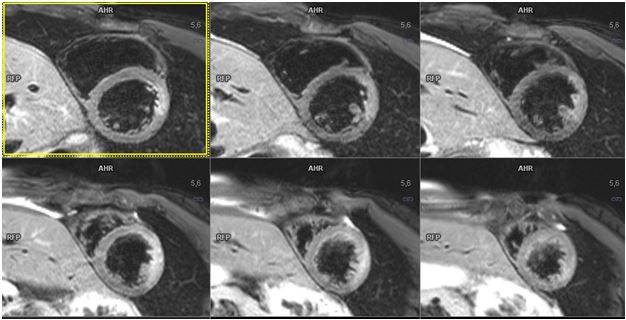
Figure 3 Heart MRI: assesment of myocardial infarction risk area – area of tissue edema (the investigation was performed in RRPC “Cardiology”)
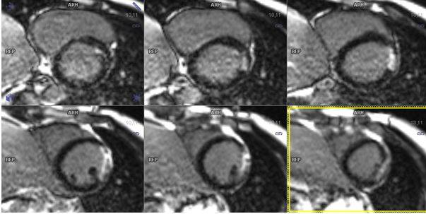
Figure 4 Heart MRI: assesment of myocardial viability using delayed Gd accumulation containing contrast medium (the investigation was performed in RRPC “Cardiology”)
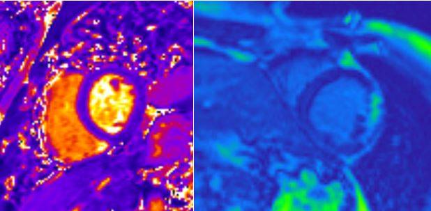
Figure 5 Heart MRI: Т12 and Т1-mapping – МР-relaxometry (the investigation was performed in RRPC “Cardiology”)
Program organ-specific analysis of vascular bed with estimate indices of the extent/length of stenoses and aneurismal dilatations, multiplanar and 3D reconstructions; SWI scanning sequence – Susceptibility weighted imaging (magnetic susceptibility) allowing for the visualization of cerebral parenchymal microhemorrhages and subsequent outcomes with highest sensitivity available at the present time; ASL scanning sequence utilized water in arterial blood as endogenous contrast medium to assess cerebral perfusion noninvasively.
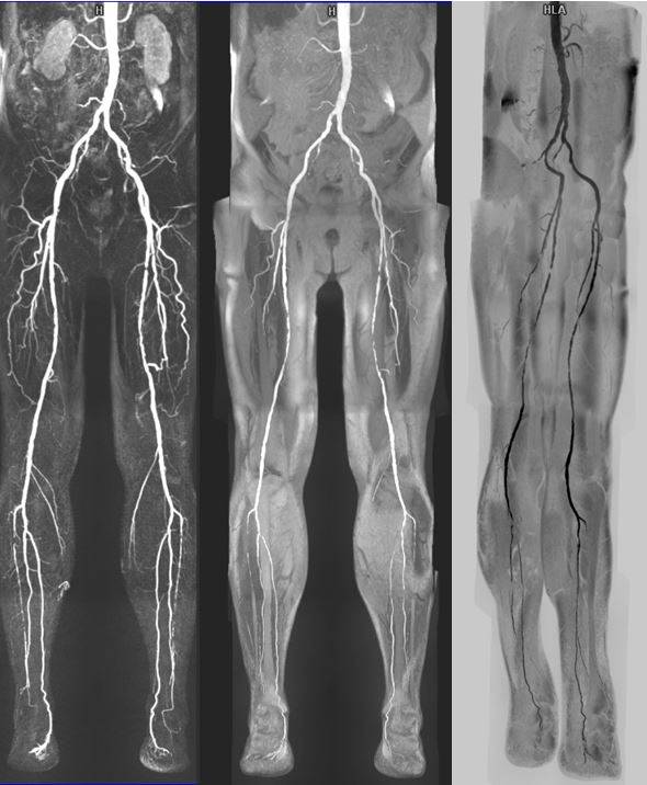
Figure 6 Contrast МР-angiography of lower limbs using Tim + Dot + Trueform technology (the investigation was performed in RRPC “Cardiology”)
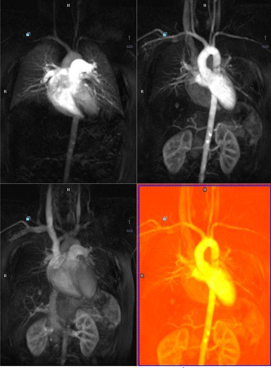
Figure 7: Dynamic contrast MRI-angiography of mediastinal vessels and abdomen (the investigation was performed in RRPC “Cardiology”)
The above-mentioned methods obtain accurate diagnostic images due to additional scanning sequences and analytical applications, maximizing diagnostic capabilities of the equipment in clinical and research application.